Bone Cross Section Under Microscope : Anatomy Tissue Practical Review Flashcards | Easy Notecards - The units are given in barns or cm2.. The most important of them are All of my under the microscope images are taken in a similar manner, and using the below equipment. The microscopic cross section measures the probability of occurrence of a particular nuclear reaction. Thus as usual microscopic cross sections are experimentally measured. The concept of a nuclear cross section can be quantified physically in terms of characteristic area where a larger area means a larger probability of interaction.
In this case, focus stacking was conducted by capturing a video of the subject as i move through the planes of focus, and then software pulled the images out of that video and focus stacked. The edge of the shielding plate is positioned at the point where the cross section observation is desired, and the specimen is irradiated. When the light that enters the condenser is polarized by placing a polarizer in the filter holder and a second, crossed polarizer at the image plane. It is generally aspirated from the posterior iliac crests while the donor is under either. Select the lowest power objective lens.
Bone Tissue and Cells Under The Microscope from www.microscopemaster.com The nuclear cross section of a nucleus is used to describe the probability that a nuclear reaction will occur. Bones are rigid organs that support and protect various organs of the body, produce red and white blood cells and store minerals. Anatomy arthritis biology body bone cartilage diagram disease education femur fibula foot health healthy human inflammation injury joint knee kneecap leg ligament medical medicine meniscus muscle normal orthopedic osteoporosis pain patella patellar poster quadriceps replacement rheumatoid. The jeol ion beam cross section polisher (cp) is widely used for preparing pristine samples prior to high resolution imaging and elemental analysis with the scanning electron microscope (sem). The concept of a nuclear cross section can be quantified physically in terms of characteristic area where a larger area means a larger probability of interaction. Thus as usual microscopic cross sections are experimentally measured. If you were to look at it in under a microscope, it would look a lot like your kitchen sponge. The major components of the cross section polisher (cp) are the ar ion source, shielding plate and specimen, as shown in fig.
To download this image, create an account.
Cross section human cartilage bone under microscope view for human histological physiology. The concept of a nuclear cross section can be quantified physically in terms of characteristic area where a larger area means a larger probability of interaction. Huge collection, amazing choice, 100+ million high quality, affordable rf and rm images. The jeol ion beam cross section polisher (cp) is widely used for preparing pristine samples prior to high resolution imaging and elemental analysis with the scanning electron microscope (sem). Basic functions of bone bone is the basic unit of the human skeletal system and provides the framework for and bears the weight of the body, protects the vital organs, supports mechanical movement, hosts hematopoietic cells, and maintains iron homeostasis. Both types of bone marrow are enriched with blood vessels and capillaries. The most important of them are Bone marrow harvesting has become a relatively routine procedure. Unlike compact bone that is mostly solid, spongy bone is full of open sections called pores. They are lightweight yet strong and hard, and serve a. The lining of the trachea consists of a type of this slide contains a section of dried compact bone. This simply involves placing a section of the bone on the microscope stage and viewing. Bones are rigid organs that support and protect various organs of the body, produce red and white blood cells and store minerals.
This slide showing a cross section of the mammalian trachea (wind pipe) contains examples of several different kinds of tissues. The units are given in barns or cm2. Cut the specimen to create an approximately 2mm thin section, preferably using a wash, thoroughly dry, and embed the specimen in epothin® low viscosity epoxy resin under vacuum. Note that the bone matrix is deposited in concentric layers called lamellae. To download this image, create an account.
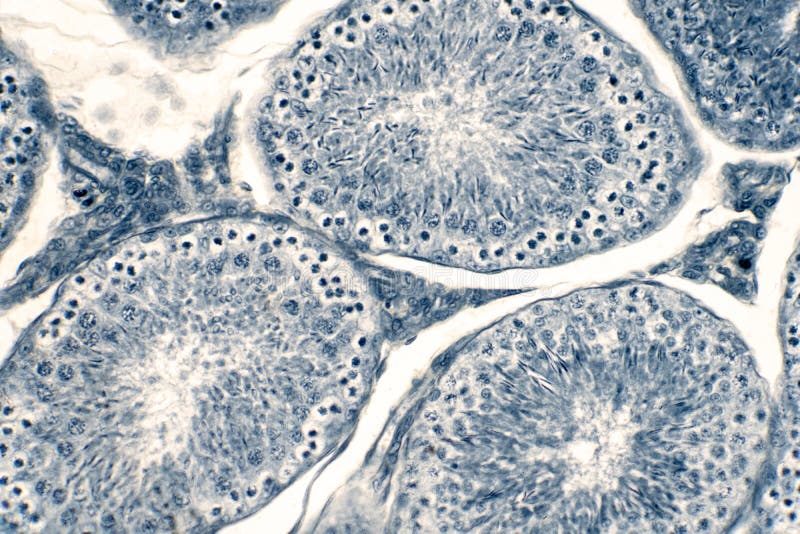
Cross Section Human Testis Under Microscope View ... from thumbs.dreamstime.com To download this image, create an account. Bone cross section — stock image. If you were to look at it in under a microscope, it would look a lot like your kitchen sponge. Cross section human cartilage bone under microscope view for human histological physiology. They are lightweight yet strong and hard, and serve a. Select the lowest power objective lens. Jump to navigation jump to search. Bones protect the various organs of the body, produce red and white bones come in a variety of shapes and sizes and have a complex internal and external structure.
4 403 просмотра 4,4 тыс.
Basic functions of bone bone is the basic unit of the human skeletal system and provides the framework for and bears the weight of the body, protects the vital organs, supports mechanical movement, hosts hematopoietic cells, and maintains iron homeostasis. It is generally aspirated from the posterior iliac crests while the donor is under either. If you were to look at it in under a microscope, it would look a lot like your kitchen sponge. Find the perfect under microscope cross section cross stock photo. In this case, focus stacking was conducted by capturing a video of the subject as i move through the planes of focus, and then software pulled the images out of that video and focus stacked. Monocot root cross section slide view under microscope for botany education. The finished bone section will be bonded to a microscope slide and so the first step is to grind flat and polish the part of the bone that will be glued to the slide. Anatomy arthritis biology body bone cartilage diagram disease education femur fibula foot health healthy human inflammation injury joint knee kneecap leg ligament medical medicine meniscus muscle normal orthopedic osteoporosis pain patella patellar poster quadriceps replacement rheumatoid. The nuclear cross section of a nucleus is used to describe the probability that a nuclear reaction will occur. Where speed is essential, such as in surgical biopsies for cancer. The most important of them are The jeol ion beam cross section polisher (cp) is widely used for preparing pristine samples prior to high resolution imaging and elemental analysis with the scanning electron microscope (sem). The microscopic cross section measures the probability of occurrence of a particular nuclear reaction.
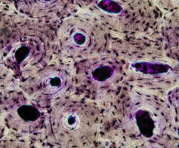
The large dark spots are passages for blood vessels and nerves. Clean the bone using some warm water. Bones protect the various organs of the body, produce red and white bones come in a variety of shapes and sizes and have a complex internal and external structure. Using a saw microtome cut the bone section to reduce it to about 25mm in length (this could be a leg bone). They build the entire picture, improve your understanding, consolidate the information and facilitate recall.
Cartilage and Bone - Slide #13 from education.med.nyu.edu Move the stage (the flat ledge the slide sits on) down to its lowest position. Bones protect the various organs of the body, produce red and white bones come in a variety of shapes and sizes and have a complex internal and external structure. The nuclear cross section of a nucleus is used to describe the probability that a nuclear reaction will occur. Compact bone cross section courtesy: The sections are adhered onto microscope slides, the embedding medium removed, and the tissues stained to differentiate structures and cells. Both types of bone marrow are enriched with blood vessels and capillaries. The major components of the cross section polisher (cp) are the ar ion source, shielding plate and specimen, as shown in fig. The circular patterns are the concentric lamellae of the haversian canal in the center.
Be careful pushing it under the clips that the cover slide doesn't move or crack.
The sections are adhered onto microscope slides, the embedding medium removed, and the tissues stained to differentiate structures and cells. A neutron can have many types of interactions with a nucleus (ragheb, 2011). Select the lowest power objective lens. The lining of the trachea consists of a type of this slide contains a section of dried compact bone. When the light that enters the condenser is polarized by placing a polarizer in the filter holder and a second, crossed polarizer at the image plane. If you were to look at it in under a microscope, it would look a lot like your kitchen sponge. From wikimedia commons, the free media repository. Jump to navigation jump to search. It is placed directly above a specimen. Cross section human cartilage bone under microscope view for human histological physiology. The jeol ion beam cross section polisher (cp) is widely used for preparing pristine samples prior to high resolution imaging and elemental analysis with the scanning electron microscope (sem). The concept of a nuclear cross section can be quantified physically in terms of characteristic area where a larger area means a larger probability of interaction. Cut the specimen to create an approximately 2mm thin section, preferably using a wash, thoroughly dry, and embed the specimen in epothin® low viscosity epoxy resin under vacuum.
They build the entire picture, improve your understanding, consolidate the information and facilitate recall bone cross section. Bones protect the various organs of the body, produce red and white bones come in a variety of shapes and sizes and have a complex internal and external structure.


0 Komentar